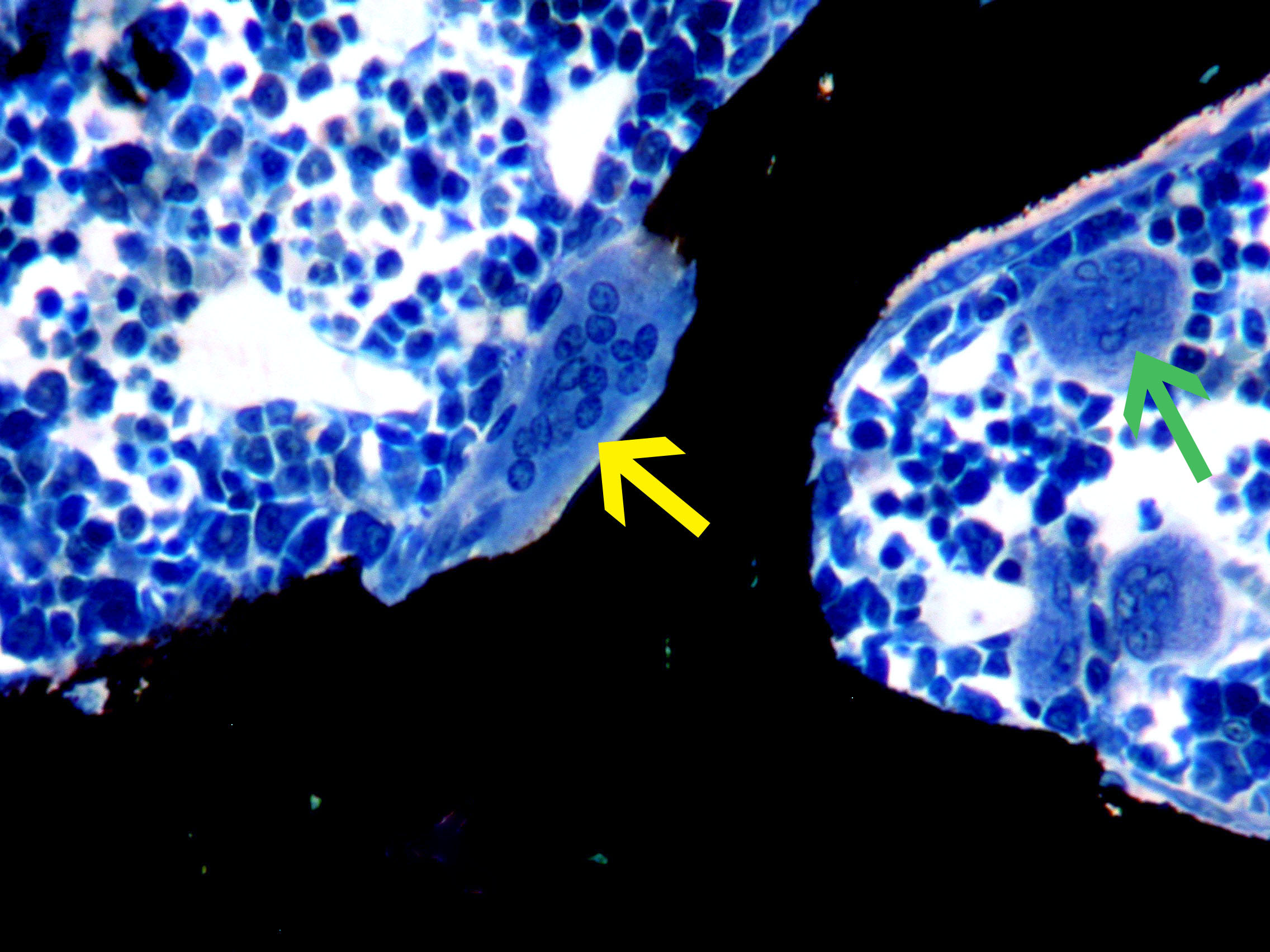
 Large Osteoclast
A large osteoclast with 10 or more nuclei adjacent to black-stained bone is indicated by the yellow arrow. This bone resorbing cell should not be confused with a megakaryocyte (green arrow).
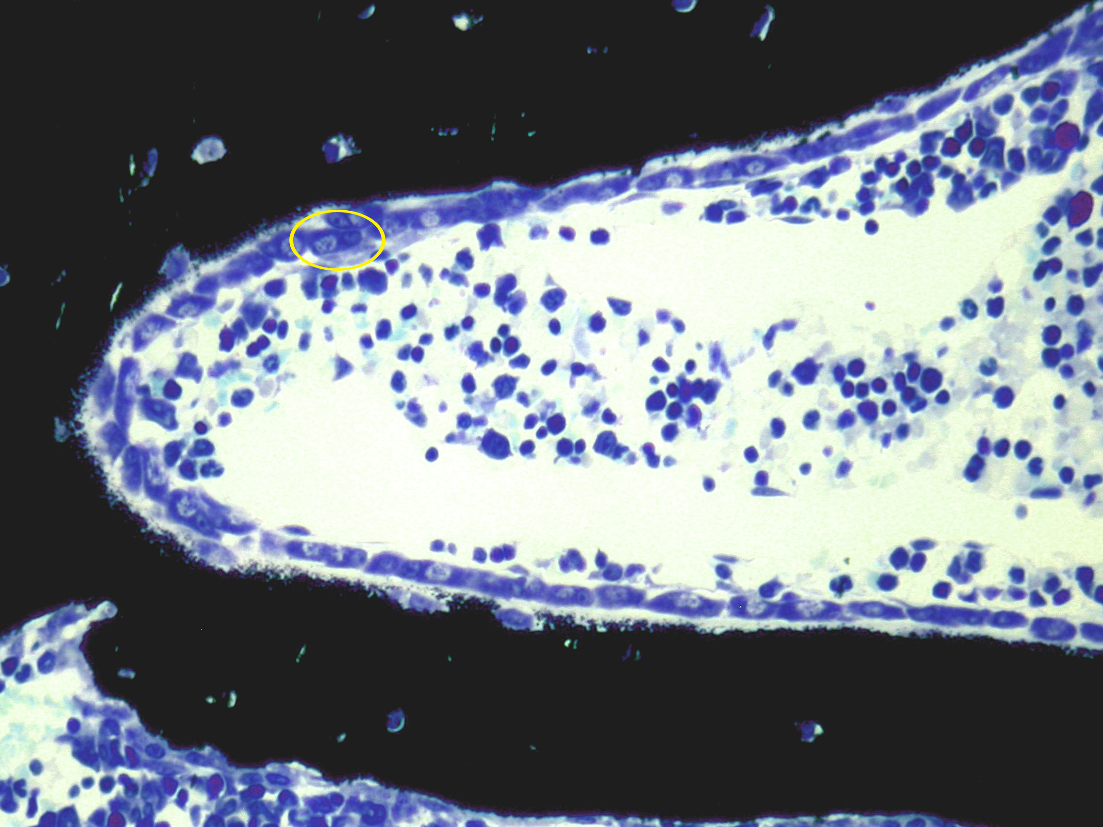 Osteoblasts in PTH-Treated Rat
The entire inner surface of black-stained bone is lined by osteoblasts. An individual osteoblast (encircled in yellow) is characterized by an eccentric nucleus and a prominent, pale-stained Golgi apparatus.
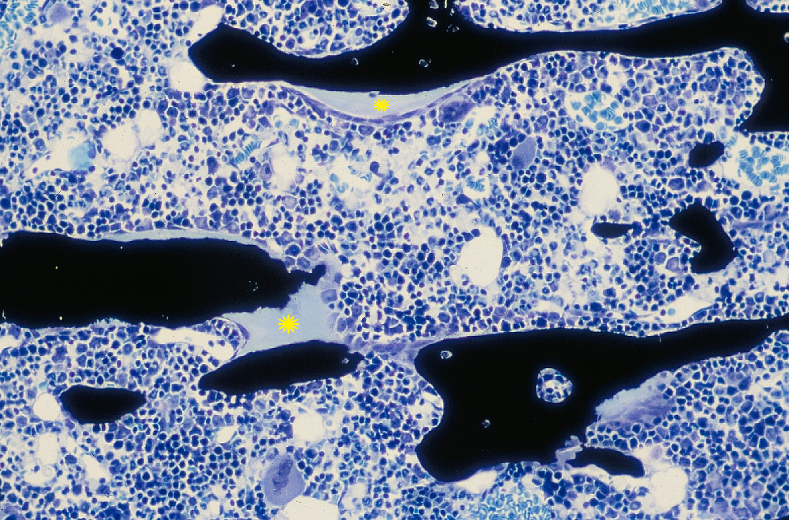 Osteoid
Osteoid is bone matrix that has not yet calcified. In this image of cancellous bone tissue, osteoid is stained light blue with a superimposed yellow starburst, and bone is stained black.
 TRAP-Stained Osteoclasts in Mouse Bone
Osteoclasts are difficult to identify in mouse bone. Therefore, it is recommended to stain for TRAP as a marker for these bone resorbing cells (yellow arrows). Since non-specific TRAP staining along bone surfaces is possible, TRAP-stained areas should be identified as osteoclasts only if nuclei are visible.
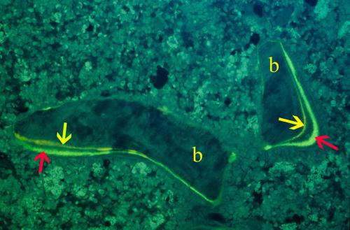 Fluorochrome Labeling of Cancellous Bone
Bone spicules (b) with double fluorochrome labels indicative of active bone formation are seen under UV illumination. The declomycin label (yellow arrows) was given at 10 days before sacrifice. The calcein label (red arrows) was given at 3 days before sacrifice.
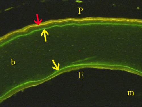 Fluorochrome Labeling of Cortical Bone
Cross section of a murine femoral shaft under UV illumination with cortical bone (b) and marrow cavity (m). The outer periosteal surface (P) exhibits a double fluorochrome label consisting of calcein (yellow arrow) and tetracycline HCl (red arrow) labels. A calcein label (yellow arrow) is also seen along the inner endocortical (E) surface.
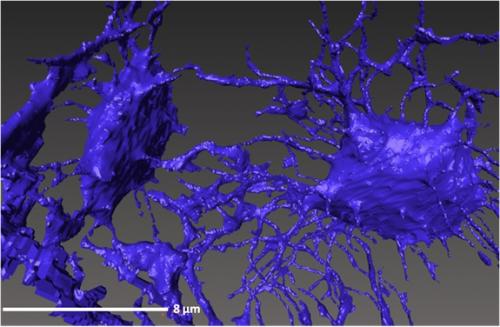 Osteocytes and Canaliculi
3D reconstruction of osteocyte lacunae connected by their canalicular network in trabecular bone from the human ilium (3D X-Ray microscope at 150 nm pixel resolution).
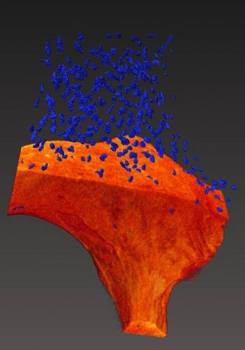 Osteocyte Lacunar Distribution
3D reconstruction of osteocyte lacunar distribution in a trabecula from a human ilium. Mineralized bone (orange) is cut away from the upper half of the image to reveal osteocyte lacunae (blue spots) within bone (3D X-Ray microscope at 0.6 micron pixel resolution).
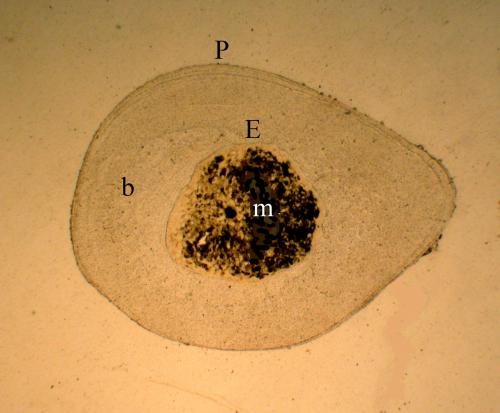 Cross Section of Rat Cortical Bone
Tibial diaphysis of an aged female rat at a standard sample site 1-2 mm proximal to the tibiofibular junction. Cortical bone (b) surrounds the marrow cavity (m) and extends between the outer, periosteal (P) surface and the inner, endocortical (E) surface
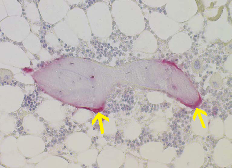
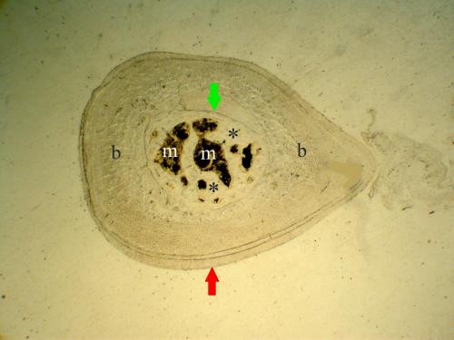 Cortical Bone in PTH-Treated Rat
Cross section of tibial diaphysis in aged female rat treated for 8 weeks with PTH. The hormone has augmented cortical bone mass (b) by stimulating deposition of layers of lamellar bone at the periosteal surface (red arrow). In addition, the marrow cavity (m) has been mostly filled by deposition of new bone (asterisks) extending from the original endocortical surface (green arrow).
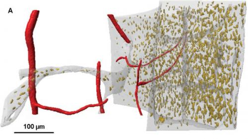 Bone Vascularity
Blood vessels (red) within the marrow cavity and cortical bone (semitransparent white) in the murine femoral mid-diaphysis. Osteocyte lacunae (yellow ellipsoids) are also visible. Vasculature was filled with a barium sulfate suspension in vivo.
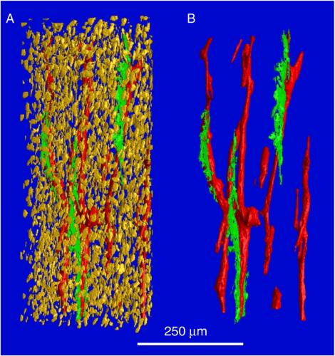 Microcracks in Cortical Bone
Canal network (red tubes), osteocyte lacunae (yellow ellipsoids), and microcracks (green planes) in the murine femoral mid-diaphysis. Osteocyte lacunae are removed in "B".
